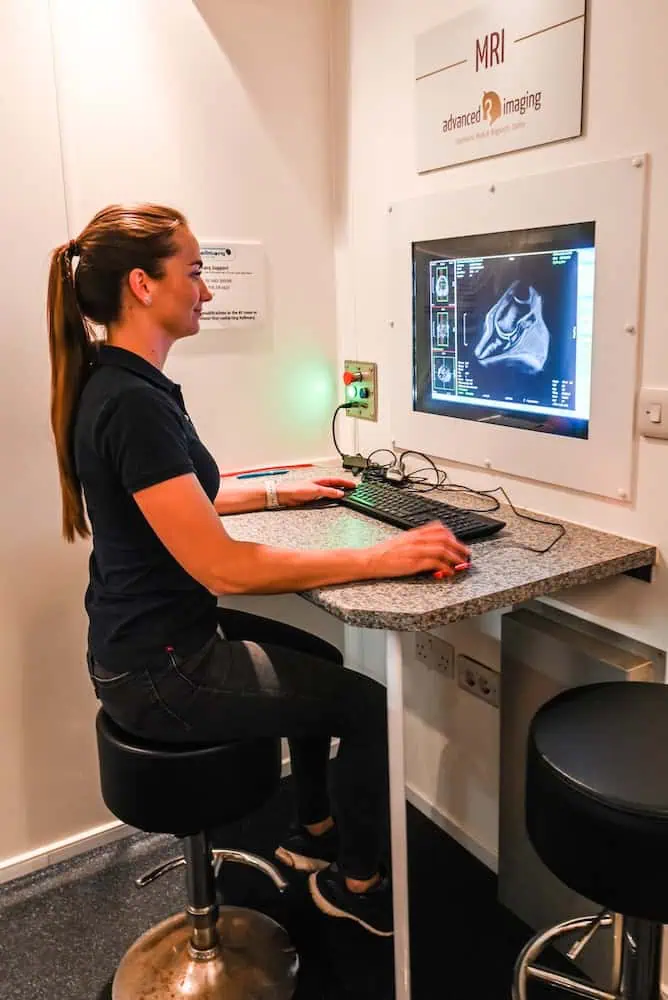
At the Sporthorse Medical Diagnostic Centre (SMDC), we perform MRI examinations using a Hallmarq standing MRI system specifically designed for horses. This allows the procedure to be carried out safely under light sedation, without the need for general anesthesia.
How does MRI work?
Magnetic Resonance Imaging (MRI) uses a powerful magnet and radio waves to produce detailed images of soft tissues and bone. Hydrogen atoms in the body are stimulated to vibrate and emit signals, which are captured by an antenna (coil) and converted into 3D images using advanced software. This allows tendon, ligament, and bone structures to be evaluated in great detail.
Indications for MRI
MRI is particularly valuable for visualizing:
- Tendon and Ligament Injuries
- Navicular Apparatus Disorders
- Joint Disorders
- Bone Bruising or Inflammation
- Causes of Unexplained Lameness after radiographs and ultrasound
We can scan the lower limb up to the carpus in the forelimb and up to the hock in the hindlimb, provided the horse remains calm and still.
Because MRI focuses on a relatively small area, it is important to perform nerve or joint blocks beforehand to accurately localize the source of pain. This ensures that we scan the correct region.
The MRI Examination
Practical Information:
- The examination takes 45 to 90 minutes per region.
- Shoes must be removed beforehand to prevent image distortion. This can be done at SMDC.
- If a farrier is present, the shoes can be reapplied after the examination.
- After the examination, the horse can usually return home immediately, but it may also stay at SMDC until the results are available.
- All images are evaluated by our own European Specialist in Veterinary Diagnostic Imaging (ECVDI).
- Within 48 hours, you will receive a written report with the findings, along with a verbal consultation with the owner and/or referring veterinarian.