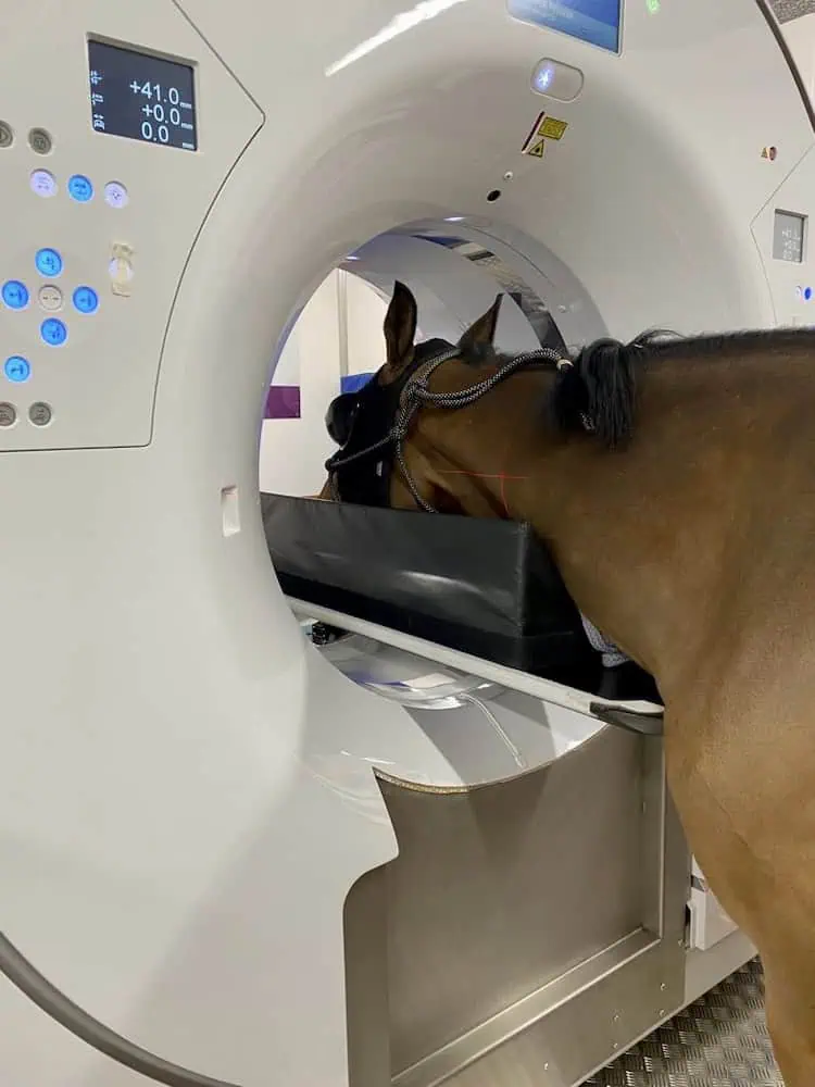
At the Sporthorse Medical Diagnostic Centre, we have an advanced Qalibra Dual Energy CT scanner with a 90-centimeter diameter—the largest available—specifically designed for horses. This system produces three-dimensional images with exceptional resolution, allowing even subtle abnormalities in bone, joint, cartilage, and surrounding tissues to be visualized.
Full-body scanning possible
Thanks to the ability to perform standing scans under light sedation, we can image the horse’s head, neck up to C3–C5, forelimbs up to the carpus, and hind legs up to the hock. For more extensive examinations, scans can be performed under general anesthesia. In this setting, nearly the entire body can be visualized, including the lower part of the neck, the thoracic transition, shoulder, elbow, and stifle.
Head and Neck
Used to diagnose conditions such as dental problems, jaw fractures, sinus infections, nerve compression, and cervical vertebral abnormalities. Because of the complex anatomy in this region, these structures are often difficult to evaluate accurately on standard X-rays.
Lower Limb up to the Hock and Carpus
Evaluation of bone and cartilage injuries, fractures, and soft tissue structures.
Complex Bone Abnormalities
Subtle fractures, cysts, or bone changes that are not clearly visible on radiographs.
Joint Disorders
Damage to cartilage or joint surfaces.
Atypical Lameness
When the cause cannot be visualized using X-ray or ultrasound.
Anesthesia is performed in consultation with a European Specialist in Anesthesiology, and the horse is safely assisted during recovery.
Myelogram
In specific cases, we also perform a myelogram, in which contrast fluid is injected into the spinal canal to visualize the space around the spinal cord and nerves. This technique is used, for example, in horses with neurological symptoms or suspected cervical stenosis or nerve compression.
Advantages of CT over radiographs
CT offers significant advantages over X-rays: it eliminates overlapping structures, reveals three-dimensional relationships, and provides a much more accurate view of dental issues, fractures, arthritic changes, and other conditions.
All images are evaluated by our own ECVDI Diplomate (European Specialist in Veterinary Diagnostic Imaging). Results are discussed with the owner or referring veterinarian within 48 hours, including a written report.
Have your horse examined by SMDC?
Together with top specialists in their fields, we ensure thorough examination and treatment.
Contact us to schedule an appointment.