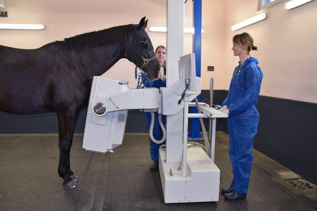
Advanced Imaging
SMDC Advanced Imaging is a collaboration between SMDC and topsporthorse veterinarians Dr.Frank van Hoeck and Dr.Willem Verhaeghe. Utilising our advanced imaging techniques, MRI and Scintigraphy, we are able to examine the standing horse in greater detail than ever before. These standing techniques do not require anesthesia so return examinations are not an issue which allows for excellent follow-up recommendations.The interpretation and reports are done by various collaborating international radiologists to ensure excellence.
Scintigraphy
Equine scintigraphy (bone scan) involves injection of a radioisotope, (technetium-99m) which is linked to a compound called bisphosphonate-MDP. Within a couple of hours this combined compound shows up in areas of the horse’s skeleton where there is increased activity of the bone, which may indicate lesions. Because the radioactivity of the compound gradually decreases, radiation is emitted from the horse’s body. This radiation (gamma radiation) is captured by a gamma camera. Through computer software, an image is acquired where abnormalities in bone activity can be detected as so called “hot spots”.
Scintigraphy is a very sensitive technique which can detect bony changes much earlier than radiography (sometimes even years before radiographic changes can be seen). Our gamma camera is designed to enable quick and efficient acquisition of scintigraphic images of any area of the axial and appendicular skeleton in the standing horse, meaning no general anaesthesia is required. Dr.Bergman is the founder of equine scintigraphy in The Netherlands (since 2003). Due to this, SMDC offers a lot of expertise and experience in performing scintigraphy and analyzing the results. SMDC is currently the only clinic in the Netherlands that has the newest and most up to date scintigraphic equipment.
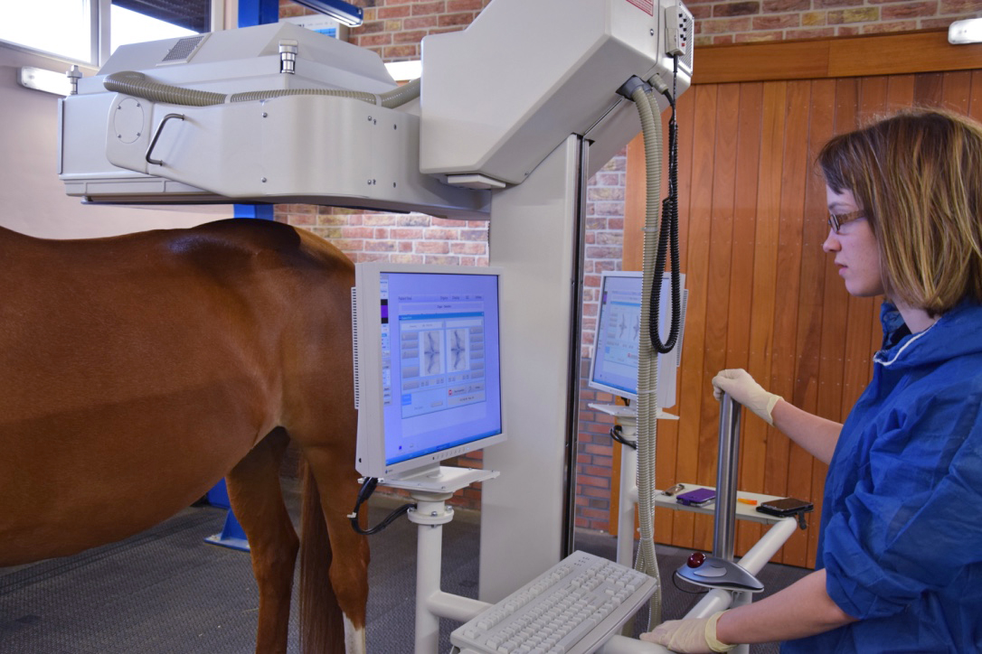
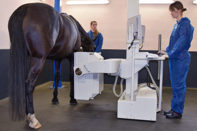
MRI
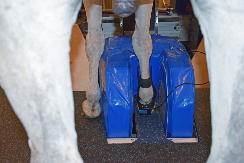
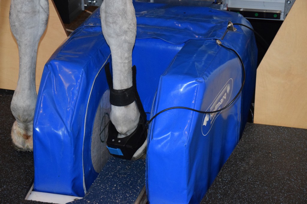
MRI stands for Magnetic Resonance Imaging. SMDC Advanced Imaging has a high quality standing MRI for horses. MRI produces highly detailed quality images that can show any abnormalities in tendons, ligaments, bone and other structures. In the front legs we are able to image from the foot to the carpus and in the hind limb from the foot to the hock.
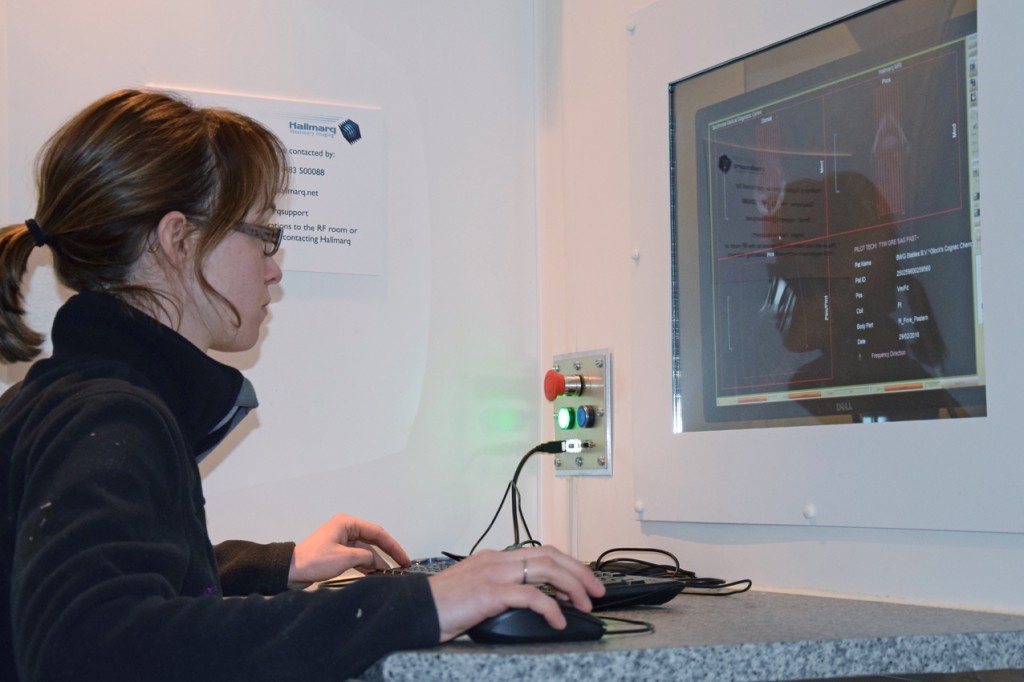
Every part of the body contains lots of hydrogen atoms (protons). These are the atoms that are responsible for getting us the MRI image with the help of a very strong magnet and radiowaves. These radiowaves are captured by a coil and via computer software get translated into a detailed image. Different structures can be imaged via different radiowaves and different sequences. MRI is very versatile in the ability to provide images sliced in many planes, and is capable of producing 3-dimensional images in a variety of orientations. These highly detailed images get interpreted by collaborating board certified radiologists.